DOE Joint Genome Institute

  • COVID-19
  • About Us
  • Contact Us
  • Our Science
    • DOE Mission Areas
    • Bioenergy Research Centers
    • Science Programs
    • Science Highlights
    • Scientists
    Data yielded from RIViT-seq increased the number of sigma factor-gene pairs confirmed in Streptomyces coelicolor from 209 to 399. Here, grey arrows denote previously known regulation and red arrows are regulation identified by RIViT-seq; orange nodes mark sigma factors while gray nodes mark other genes. (Otani, H., Mouncey, N.J. Nat Commun 13, 3502 (2022). https://doi.org/10.1038/s41467-022-31191-w)
    Streamlining Regulon Identification in Bacteria
    Regulons are a group of genes that can be turned on or off by the same regulatory protein. RIViT-seq technology could speed up associating transcription factors with their target genes.

    More

    (PXFuel)
    Designer DNA: JGI Helps Users Blaze New Biosynthetic Pathways
    In a special issue of the journal Synthetic Biology, JGI scientific users share how they’ve worked with the JGI DNA Synthesis Science Program and what they’ve discovered through their collaborations.

    More

    A genetic element that generates targeted mutations, called diversity-generating retroelements (DGRs), are found in viruses, as well as bacteria and archaea. Most DGRs found in viruses appear to be in their tail fibers. These tail fibers – signified in the cartoon by the blue virus’ downward pointing ‘arms’— allow the virus to attach to one cell type (red), but not the other (purple). DGRs mutate these ‘arms,’ giving the virus opportunities to switch to different prey, like the purple cell. (Courtesy of Blair Paul)
    A Natural Mechanism Can Turbocharge Viral Evolution
    A team has discovered that diversity generating retroelements (DGRs) are not only widespread, but also surprisingly active. In viruses, DGRs appear to generate diversity quickly, allowing these viruses to target new microbial prey.

    More

  • Our Projects
    • Search JGI Projects
    • DOE Metrics/Statistics
    • Approved User Proposals
    • Legacy Projects
    Photograph of a stream of diatoms beneath Arctic sea ice.
    Polar Phytoplankton Need Zinc to Cope with the Cold
    As part of a long-term collaboration with the JGI Algal Program, researchers studying function and activity of phytoplankton genes in polar waters have found that these algae rely on dissolved zinc to photosynthesize.

    More

    This data image shows the monthly average sea surface temperature for May 2015. Between 2013 and 2016, a large mass of unusually warm ocean water--nicknamed the blob--dominated the North Pacific, indicated here by red, pink, and yellow colors signifying temperatures as much as three degrees Celsius (five degrees Fahrenheit) higher than average. Data are from the NASA Multi-scale Ultra-high Resolution Sea Surface Temperature (MUR SST) Analysis product. (Courtesy NASA Physical Oceanography Distributed Active Archive Center)
    When “The Blob” Made It Hotter Under the Water
    Researchers tracked the impact of a large-scale heatwave event in the ocean known as “The Blob” as part of an approved proposal through the Community Science Program.

    More

    A plantation of poplar trees. (David Gilbert)
    Genome Insider podcast: THE Bioenergy Tree
    The US Department of Energy’s favorite tree is poplar. In this episode, hear from ORNL scientists who have uncovered remarkable genetic secrets that bring us closer to making poplar an economical and sustainable source of energy and materials.

    More

  • Data & Tools
    • IMG
    • Data Portal
    • MycoCosm
    • PhycoCosm
    • Phytozome
    • GOLD
    HPCwire Editor's Choice Award (logo crop) for Best Use of HPC in the Life Sciences
    JGI Part of Berkeley Lab Team Awarded Best Use of HPC in Life Sciences
    The HPCwire Editors Choice Award for Best Use of HPC in Life Sciences went to the Berkeley Lab team comprised of JGI and ExaBiome Project team, supported by the DOE Exascale Computing Project for MetaHipMer, an end-to-end genome assembler that supports “an unprecedented assembly of environmental microbiomes.”

    More

    With a common set of "baseline metadata," JGI users can more easily access public data sets. (Steve Wilson)
    A User-Centered Approach to Accessing JGI Data
    Reflecting a structural shift in data access, the JGI Data Portal offers a way for users to more easily access public data sets through a common set of metadata.

    More

    Phytozome portal collage
    A More Intuitive Phytozome Interface
    Phytozome v13 now hosts upwards of 250 plant genomes and provides users with the genome browsers, gene pages, search, BLAST and BioMart data warehouse interfaces they have come to rely on, with a more intuitive interface.

    More

  • User Programs
    • Calls for Proposals
    • Special Initiatives & Programs
    • Product Offerings
    • User Support
    • Policies
    • Submit a Proposal
    screencap from Amundson and Wilkins subsurface microbiome video
    Digging into Microbial Ecosystems Deep Underground
    JGI users and microbiome researchers at Colorado State University have many questions about the microbial communities deep underground, including the role viral infection may play in other natural ecosystems.

    Read more

    Yeast strains engineered for the biochemical conversion of glucose to value-added products are limited in chemical output due to growth and viability constraints. Cell extracts provide an alternative format for chemical synthesis in the absence of cell growth by isolating the soluble components of lysed cells. By separating the production of enzymes (during growth) and the biochemical production process (in cell-free reactions), this framework enables biosynthesis of diverse chemical products at volumetric productivities greater than the source strains. (Blake Rasor)
    Boosting Small Molecule Production in Super “Soup”
    Researchers supported through the Emerging Technologies Opportunity Program describe a two-pronged approach that starts with engineered yeast cells but then moves out of the cell structure into a cell-free system.

    More

    These bright green spots are fluorescently labelled bacteria from soil collected from the surface of plant roots. For reference, the scale bar at bottom right is 10 micrometers long. (Rhona Stuart)
    A Powerful Technique to Study Microbes, Now Easier
    In JGI's Genome Insider podcast: LLNL biologist Jennifer Pett-Ridge collaborated with JGI scientists through the Emerging Technologies Opportunity Program to semi-automate experiments that measure microbial activity in soil.

    More

  • News & Publications
    • News
    • Blog
    • Podcasts
    • Webinars
    • Publications
    • Newsletter
    • Logos and Templates
    • Photos
    A view of the mangroves from which the giant bacteria were sampled in Guadeloupe. (Hugo Bret)
    Giant Bacteria Found in Guadeloupe Mangroves Challenge Traditional Concepts
    Harnessing JGI and Berkeley Lab resources, researchers characterized a giant - 5,000 times bigger than most bacteria - filamentous bacterium discovered in the Caribbean mangroves.

    More

    In their approved proposal, Frederick Colwell of Oregon State University and colleagues are interested in the microbial communities that live on Alaska’s glacially dominated Copper River Delta. They’re looking at how the microbes in these high latitude wetlands, such as the Copper River Delta wetland pond shown here, cycle carbon. (Courtesy of Rick Colwell)
    Monitoring Inter-Organism Interactions Within Ecosystems
    Many of the proposals approved through JGI's annual Community Science Program call focus on harnessing genomics to developing sustainable resources for biofuels and bioproducts.

    More

    Coloring the water, the algae Phaeocystis blooms off the side of the sampling vessel, Polarstern, in the temperate region of the North Atlantic. (Katrin Schmidt)
    Climate Change Threatens Base of Polar Oceans’ Bountiful Food Webs
    As warm-adapted microbes edge polewards, they’d oust resident tiny algae. It's a trend that threatens to destabilize the delicate marine food web and change the oceans as we know them.

    More

News & Publications
Home › News Releases › Giant Bacteria Found in Guadeloupe Mangroves Challenge Traditional Concepts

June 23, 2022

Giant Bacteria Found in Guadeloupe Mangroves Challenge Traditional Concepts

Though visible to the naked eye, microscopy reveals unexpected complexity

Single filament of Ca. Thiomargarita magnifica (Jean-Marie Volland)

Single filament of Ca. Thiomargarita magnifica (Jean-Marie Volland)

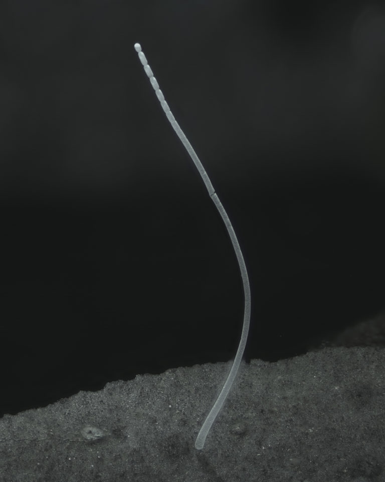
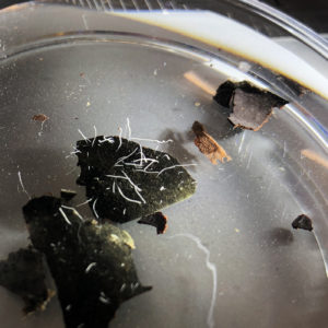
At first glance, the slightly murky waters in the tube look like a scoop of stormwater, complete with leaves, debris, and even lighter threads in the mix. But in the Petri dish, the thin vermicelli-like threads floating delicately above the leaf debris are revealed to be single bacterial cells, visible to the naked eye.

The unusual size is notable because bacteria aren’t usually visible without the assistance of microscope. “It’s 5,000 times bigger than most bacteria. To put it into context, it would be like a human encountering another human as tall as Mount Everest,” said Jean-Marie Volland, a scientist with joint appointments at the U.S. Department of Energy (DOE) Joint Genome Institute (JGI), a DOE Office of Science User Facility located at Lawrence Berkeley National Laboratory (Berkeley Lab) and the Laboratory for Research in Complex Systems (LRC) in Menlo Park, Calif. In the June 24, 2022, issue of the journal Science, Volland and colleagues, including researchers at the JGI and Berkeley Lab, LRC, and at the Université des Antilles, described the morphological and genomic features of this giant filamentous bacterium, along with its life cycle.

For most bacteria, their DNA floats freely within the cytoplasm of their cells. This newly discovered species of bacteria keeps its DNA more organized. “The big surprise of the project was to realize that these genome copies that are spread throughout the whole cell are actually contained within a structure that has a membrane,” Volland said. “And this is very unexpected for a bacterium.”

Views of the sampling sites amidst the mangroves in Guadeloupe. (Olivier Gros)

Views of the sampling sites amidst the mangroves in Guadeloupe. (Olivier Gros)

Strange Encounters in the Mangroves

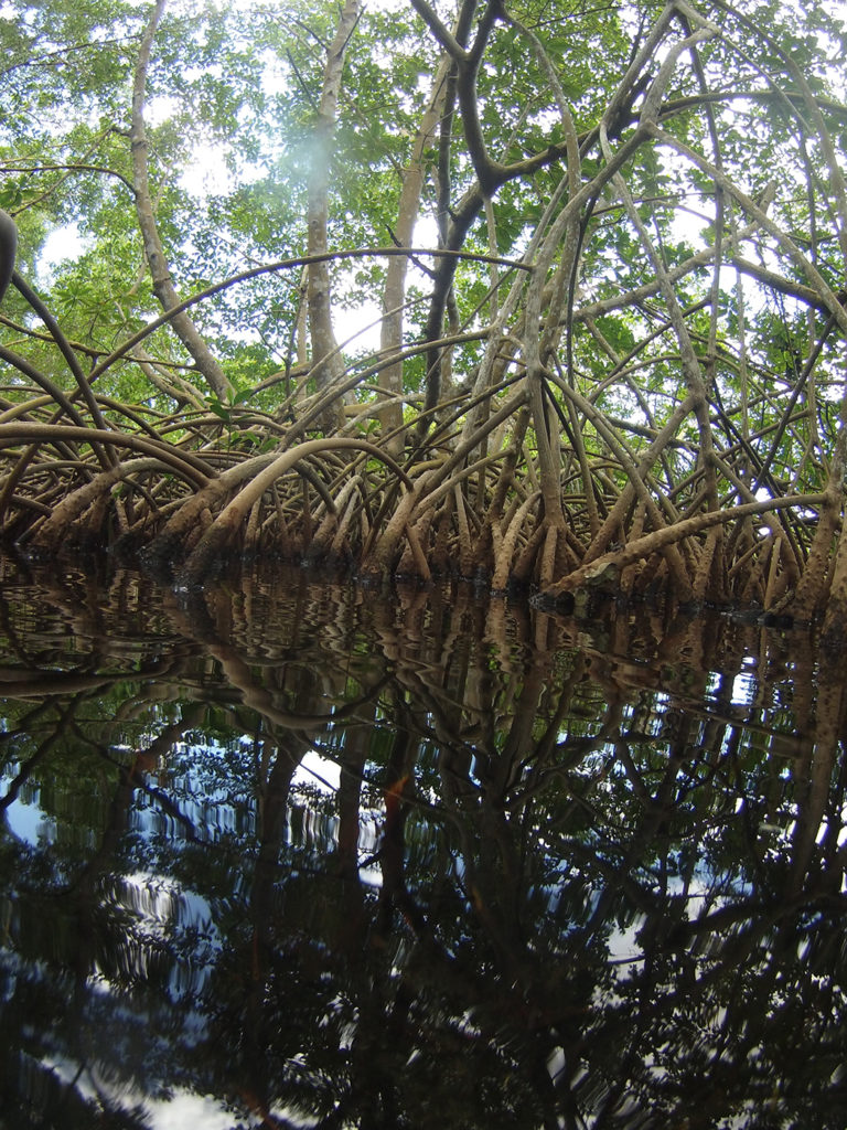
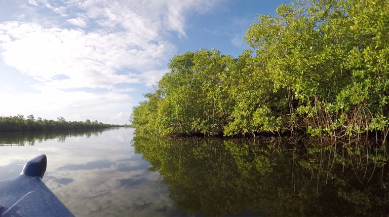
The bacterium itself was discovered by Olivier Gros, a marine biology professor at the Université des Antilles in Guadeloupe, in 2009. Gros’ research focuses on marine mangrove systems, and he was looking for sulfur-oxidizing symbionts in sulfur-rich mangrove sediments not far from his lab when he first encountered the bacteria. “When I saw them, I thought, ‘Strange,’” he said. “In the beginning I thought it was just something curious, some white filaments that needed to be attached to something in the sediment like a leaf.” The lab conducted some microscopy studies over the next couple of years, and realized it was a sulfur-oxidizing prokaryote.

Silvina Gonzalez-Rizzo, an associate professor of molecular biology at the Université des Antilles and a co-first author on the study, performed the 16S rRNA gene sequencing to identify and classify the prokaryote. “I thought they were eukaryotes; I didn’t think they were bacteria because they were so big with seemingly a lot of filaments,” she recalled of her first impression. “We realized they were unique because it looked like a single cell. The fact that they were a ‘macro’ microbe was fascinating!”

“She understood that it was a bacterium belonging to the genus Thiomargarita,” Gros noted. “She named it Ca. Thiomargarita magnifica.”

“Magnifica because magnus in Latin means big and I think it’s gorgeous like the French word magnifique,” Gonzalez-Rizzo explained. “This kind of discovery opens new questions about bacterial morphotypes that have never been studied before.”

Filaments of Ca. Thiomargarita magnifica attached to leaf debris are visible to the naked eye on a Petri dish. (Massie S. Ballon)

Filaments of Ca. Thiomargarita magnifica attached to leaf debris are visible to the naked eye on a Petri dish. (Massie S. Ballon)

Characterizing the Giant Bacterium

Volland got involved with the giant Thiomargarita bacteria when he returned to the Gros lab as a postdoctoral fellow. When he applied to the discovery-based position at the LRC that would see him working at the JGI, Gros allowed him to continue research on the project.

At the JGI, Volland began studying Ca. T. magnifica in Tanja Woyke’s Single Cells Group to better understand what this sulfur-oxidizing, carbon fixing bacterium was doing in the mangroves. “Mangroves and their microbiomes are important ecosystems for carbon cycling. If you look at the space that they occupy on a global scale, it’s less than 1% of the coastal area worldwide. But when you then look at carbon storage, you’ll find that they contribute 10-15% of the carbon stored in coastal sediments,” said Woyke, who also heads the JGI’s Microbial Program and is one of the article’s senior authors. The team was also compelled to study these large bacteria in light of their potential interactions with other microorganisms. “We started this project under the JGI’s strategic thrust of inter-organismal interactions, because large sulfur bacteria have been shown to be hot spots for symbionts”, Woyke said. “Yet the project took us into a very different direction,” she added.

Volland took on the challenge to visualize these giant cells in three dimensions and at relatively high magnification. Using various microscopy techniques, such as hard x-ray tomography, for instance, he visualized entire filaments up to 9.66 mm long and confirmed that they were indeed giant single cells rather than multicellular filaments, as is common in other large sulfur bacteria. He was also able to use imaging facilities available at Berkeley Lab, such as confocal laser scanning microscopy and transmission electron microscopy (TEM) to visualize the filaments and the cell membranes in more details. These techniques allowed him to observe novel, membrane-bound compartments that contain DNA clusters. He dubbed these organelles “pepins,” after the small seeds in fruits. DNA clusters were plentiful in the single cells.

Some of the authors of the Science publication. Left to right: Tanja Woyke, Jean-Marie Volland, Olivier Gros, Silvina Gonzalez-Rizzo and Shailesh Date. (Background image by Hugo Bret; Composite by Susan Brand/Berkeley Lab)

Some of the authors of the Science publication. Left to right: Tanja Woyke, Jean-Marie Volland, Olivier Gros, Silvina Gonzalez-Rizzo and Shailesh Date. (Background image by Hugo Bret; Composite by Susan Brand/Berkeley Lab)

The team learned about the cell’s genomic complexity. As Volland noted, “The bacteria contain three times more genes than most bacteria and hundreds of thousands of genome copies (polyploidy) that are spread throughout the entire cell.” The JGI team then used single cell genomics to analyze five of the bacterial cells on the molecular level. They amplified, sequenced and assembled the genomes. In parallel, Gros’ lab also used a labeling technique known as BONCAT to identify areas involved in protein-making activities, that confirmed that the entire bacterial cells were active.

“This project has been a nice opportunity to demonstrate how complexity has evolved in some of the simplest organisms,” said Shailesh Date, founder and CEO of LRC, and one of the article’s senior authors. “One of the things we’ve argued is that there is need to look at and study biological complexity in much more detail than what is being done currently. So organisms that we think are very, very simple might have some surprises.”

The LRC provided funding for Volland through grants from the John Templeton Foundation and the Gordon and Betty Moore Foundation. “This groundbreaking discovery highlights the importance of supporting fundamental, creative research projects to advance our understanding of the natural world,” added Sara Bender of the Gordon and Betty Moore Foundation. “We look forward to learning how the characterization of Ca. Thiomargarita magnifica challenges the current paradigm of what constitutes a bacterial cell and advances microbial research.”

A view of the mangroves from which the giant bacteria were sampled in Guadeloupe. (Hugo Bret)

A view of the mangroves from which the giant bacteria were sampled in Guadeloupe. (Hugo Bret)

One Giant Bacterium, Multiple Research Questions

For the team, characterizing Ca. Thiomargarita magnifica has paved the way for multiple new research questions. Among them, is the bacterium’s role in the mangrove ecosystem. “We know that it’s growing and thriving on top of the sediment of mangrove ecosystem in the Caribbean,” Volland said. “In terms of metabolism, it does chemosynthesis, which is a process analogous to photosynthesis for plants.” Another outstanding question is whether the new organelles named pepins played a role in the evolution of the Thiomargarita magnifica extreme size, and whether or not pepins are present in other bacterial species. The precise formation of pepins and how molecular processes within and outside of these structures occur and are regulated  also remain to be studied.

Gonzalez-Rizzo and Woyke both see successfully cultivating the bacteria in the lab as a way to get some of the answers. “If we can maintain these bacteria in a laboratory setting, we can use techniques that are not feasible right now”, Woyke said. Gros wants to look at other large bacteria. “You can find some TEM pictures and see what look like pepins so maybe people saw them but did not understand what they were. That will be very interesting to check, if the pepins are already present everywhere.”

Researchers from the National Museum of Natural History (France), Sorbonne University (France) and Cornell University were also involved in this work. Other authors on the paper are Tomáš Tyml, Natalia Ivanova, Frederik Schulz, Danielle Goudeau, Nathalie H. Elisabeth, Nandita Nath, Daniel Udwary, Rex R. Malmstrom, Chantal Guidi-Rontani, Susanne Bolte-Kluge, Karen M. Davies, Maïtena R. Jean, Jean-Louis Mansot, Nigel J. Mouncey, and Esther R. Angert.


Publication:

  • Volland JM*, Gonzalez-Rizzo S*, Gros O* et al. A centimeter-long bacterium with DNA contained in metabolically active membrane-bound organelles. Science. 2022 June. doi: 10.1126/science.abb3634

*co-first authors


Byline: Massie S. Ballon

Share this:

  • Click to share on Facebook (Opens in new window)
  • Click to share on LinkedIn (Opens in new window)
  • Click to share on Pinterest (Opens in new window)
  • Click to share on Twitter (Opens in new window)
  • Click to print (Opens in new window)

The U.S. Department of Energy Joint Genome Institute, a DOE Office of Science User Facility at Lawrence Berkeley National Laboratory, is committed to advancing genomics in support of DOE missions related to clean energy generation and environmental characterization and cleanup. JGI provides integrated high-throughput sequencing and computational analysis that enable systems-based scientific approaches to these challenges. Follow @jgi on Twitter.

DOE’s Office of Science is the largest supporter of basic research in the physical sciences in the United States, and is working to address some of the most pressing challenges of our time. For more information, please visit science.energy.gov.

Filed Under: News Releases

More topics:

  • COVID-19 Status
  • News
  • Science Highlights
  • Blog
  • Webinars
  • CSP Plans
  • Featured Profiles

Related Content:

Busting the Unbreakable Lignin

Pictured is a micrograph of Neocallimastix californiae.

Tracing the Evolution of Shiitake Mushrooms

A vertical tree stump outdoors with about a dozen shiitake mushrooms sprouting from its surface.

JGI announces final round of 2022 Functional Genomics awardees

Digital ID card with six headshots reads: Congratulations to our 2022 Function Genomics recipients!

Introducing New Members of the JGI User Executive Committee

incoming 2023 UEC members

JGI at 25: Mapping Switchgrass Traits with Common Gardens

Aerial photo of the switchgrass diversity panel late in the 2020 season at the Kellogg Biological Station in Michigan. (Robert Goodwin)

JGI Contributes Nine to 2022 Highly Cited Researchers List

Nine headshots, one for each researcher, laid out beside a purple ribbon reading, "Home to Highly Cited Researchers 2022 Clarivate"
  • Careers
  • Contact Us
  • Events
  • User Meeting
  • MGM Workshops
  • Internal
  • Disclaimer
  • Credits
  • Policies
  • Emergency Info
  • Accessibility / Section 508 Statement
  • Flickr
  • LinkedIn
  • RSS
  • Twitter
  • YouTube
Lawrence Berkeley National Lab Biosciences Area
A project of the US Department of Energy, Office of Science

JGI is a DOE Office of Science User Facility managed by Lawrence Berkeley National Laboratory

© 1997-2023 The Regents of the University of California